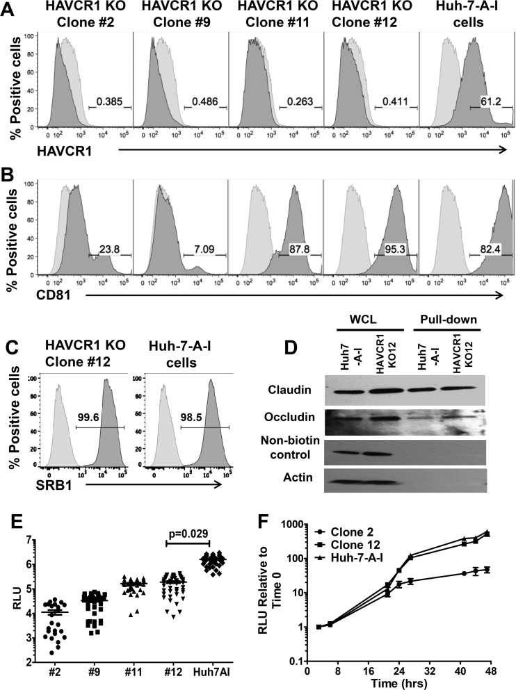
FIG 5.
Complete silencing of HAVCR1 expression in Huh7.5 cells significantly reduces HCVcc infection. Surface expression levels of receptors on Huh7.5 cells, Huh7-A-I cells, and different clones of Huh7-A-I HAVCR1 knockout (KO) cells were assessed by FACS using MAbs specific to HAVCR1 (REA383) (A), CD81 (JS-81) (B), and SRB1 (m1B9) (C). Data for positive cells are shown in dark gray, and data for mouse PE-isotype control are shown in light gray. Data are representative of three independent experiments. (D) Western blot detection of claudin and occludin after cell surface biotinylation of Huh7-A-I and clone 12 KO cells. Whole-cell lysate (WCL) is cell lysate prior to pulldown treatment. Nonbiotinylated control represents cells treated exactly as for the biotinylated cells but without the addition of EZ-Link sulfo-NHS-LCLC-biotin for cell surface biotinylation. Anticlaudin antibody was used for detection and shows the specificity of the NeutrAvidin beads for precipitation. Antiactin antibody was used for detection of actin, a cytoplasmic protein that is not expressed on the cell surface, demonstrating biotinylation of surface proteins only. (E) Luciferase activities in lysates from Huh7-A-I HAVCR1 KO clones and parent cell line at 72 h postinfection with JFH1-Nanoluc. Data were obtained from 3 independent experiments, and each experiment was carried out using 12 replicates for each cell clone. Each symbol represents an individual well from the 3 independent experiments. Horizontal bars represent the mean luciferase activities. P values were calculated using the mean values from each of the 3 independent experiments. (F) Time course of luciferase expression relative to the expression at 3 h posttransfection in Huh7-A-I HAVCR1 KO clones 2 and 12 and parent cells following transfection with JFH1-Nanoluc RNA transcribed in vitro. Data are the mean values from 6 replicates at each time point. Error bars represent standard errors of the means. Data are representative of two independent experiments. RLU, relative light units.

