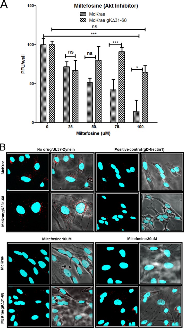
FIG 1.
Effect of an Akt inhibitor on HSV-1 entry. (A) Vero cells were treated with a series of dilutions of the Akt inhibitor miltefosine (labeled Akt inhibitor) for 15 min and then adsorbed with McKrae and McKrae gKΔ31-68 (100 PFU) for 1 h at 4°C. After incubation at 37°C for 48 h, the plaques (the number of PFU) were counted using crystal violet staining. *, P < 0.05 between McKrae and gKΔ31-68 viruses; ***, P < 0.001 versus no-drug-treated control; ns, no significance versus no-drug-treated control. Statistical comparison was conducted by GraphPad Prism software using ANOVA with a post hoc t test with the Bonferroni adjustment. Bars represent 95% confidence intervals about the means. (B) SK-N-SH cells were treated with miltefosine for 15 min and infected with McKrae or McKrae gKΔ31-68 (MOI = 10) for 1 h at 37°C, and the proximity ligation assay (PLA) was performed. Confocal microscopy was used to detect bright red spots, which indicate an interaction between two proteins after drug treatment, at a magnification of ×63 with oil immersion. The interaction between UL37 (capsid protein) and dynein (cellular protein) was used as a measure of entry of the virus. The interaction between gD and nectin-1 was used as a positive PLA control in this experiment. DAPI was used to stain the nuclei of the cells.

