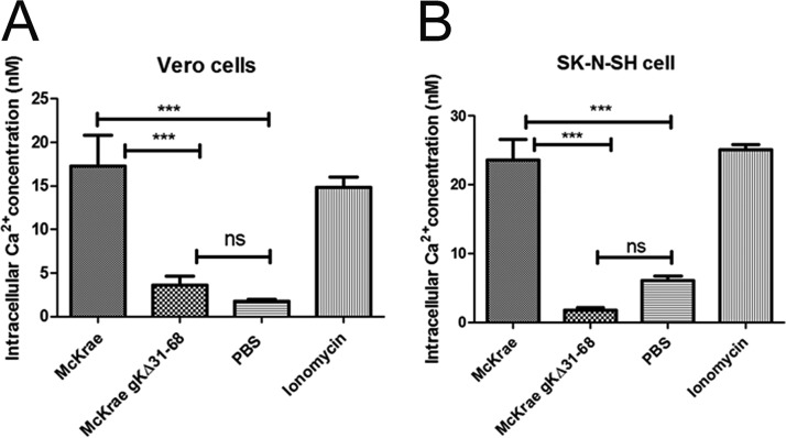
FIG 3.
HSV-1 triggers intracellular calcium release. Vero cells (A) and SK-N-SH cells (B) were grown in a 96-well black clear-bottom plate and treated with Fura-2AM (25 μM) for 30 min at 37°C. After washing, the cells were adsorbed with purified McKrae and McKrae gKΔ31-68 (both at an MOI of 10) at 4°C for 1 h and then shifted to 37°C to measure the absorbance every minute over an hour. The ratio of bound to unbound dye (340/380 nm) was measured, the intracellular calcium concentration was calculated, and the average values are presented. Ionomycin was used as a positive control, and 1× PBS was used as a negative control. ***, P < 0.001 versus the PBS control; ns, no significance versus the PBS control. Statistical comparison was conducted by GraphPad Prism software using ANOVA with a post hoc t test with the Bonferroni adjustment. Bars represent the 95% confidence intervals about the means.

