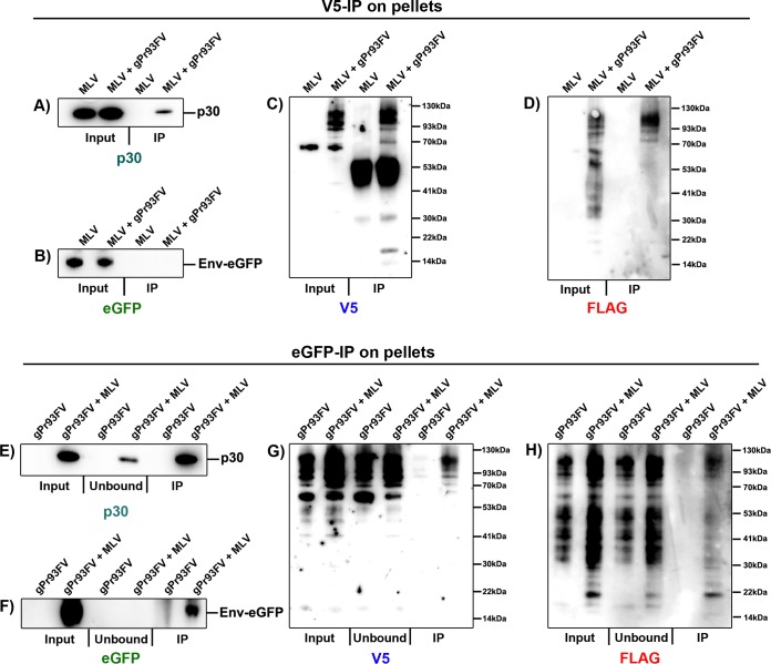
FIG 9.
Glycosylated Gag adopts a type I integral membrane protein conformation in the envelope of viral particles. 293T cells were transfected similarly to those in Fig. 8. Supernatants were pelleted through a 20% sucrose cushion by velocity sedimentation and resuspended in PBS. (A to D) The viral preparations were immunoprecipitated by use of anti-V5-conjugated magnetic beads and washed with PBS. Input fractions (lysates) and anti-V5 IP purified particles were analyzed by SDS-PAGE and probed with p30 (A), EGFP (B), V5 (C), and FLAG (D) antibodies. (E to H) Viral preparations were also immunoprecipitated using anti-EGFP-conjugated magnetic beads. Input (lysates), flowthrough (unbound), and anti-GFP (IP)-purified enveloped particles were analyzed by SDS-PAGE and probed with p30 (E), EGFP (F), V5 (G), and FLAG (H) antibodies. Data are representative of one experiment for three independent transfections.

