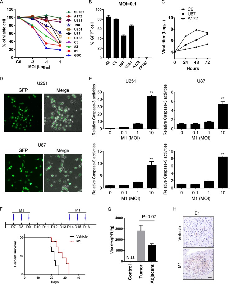
FIG 1.
Oncolytic effects of the M1 virus in vitro and in vivo. (A) A panel of glioma cell lines and primary glioma cells from patients (1 and 2) were infected with the M1 virus at 10, 0.1, 0.001, and 0 PFU/cell. Cell viabilities were determined at 48 h postinfection. MOI, multiplicity of infection. (B) Cells were infected with the M1-GFP virus at 0.1 PFU/cell, and GFP positive cells were analyzed by flow cytometry at 48 h postinfection. (C) M1 virus replication in different glioma cell lines. Cells were infected with the M1 virus (0.1 PFU/cell), and supernatants were collected to determine the viral replication. (D) Representative photographs of U87 and U251 cells infected with the M1-GFP virus (10 PFU/cell) at 72 h postinfection. (E) U87 and U251 cells were infected with the M1 virus for 24 h, and caspase-3/7/9 activities were determined. (F) Timeline of treatment for in vivo experiment and survival analysis of glioma-bearing mice. Mice were orthotopically inoculated with 3 × 10⁵ U87 cells. After 1 week, the M1 virus was injected through the tail vein. (G and H) Virus titer and expression of E1 viral protein from tissues derived from U87 orthotopic glioma model mice. N.D., not detectable. *, P < 0.05; **, P < 0.01.

