FIG 5.
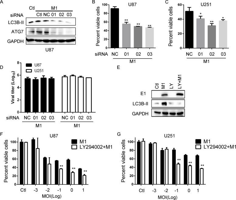
IRE1α-mediated autophagy restricts the oncolytic effects of the M1 virus. (A) LC3B detection after knockdown of ATG7. Cells were transfected with scramble RNA or siATG7 (50 nM) for 48 h. The indicated protein expression levels were determined at 8 h postinfection. (B and C) Cell viability determination after knockdown of ATG7. U87 and U251 cells were transfected with siRNA for 24 h. Cell viabilities were determined at 48 h postinfection. (D) Viral titer assay after ATG7 knockdown. Cells were treated as described for panel B, and supernatants were collected at 48 h postinfection. (E) Viral protein detection after treatment with the autophagy inhibitor LY294002. U87 cells were pretreated with LY294002 (20 μM) for 1 h and then infected with the M1 virus (1 PFU/cell). E1 and LC3B protein expression levels were determined with a Western blot. (F and G) Cell viability assay of the oncolytic effect of the M1 virus after LY294002 treatment. Cells were pretreated with LY294002 for 1 h and infected with the M1 virus for 48 h. *, P < 0.05; **, P < 0.01.

