
FIG 1.
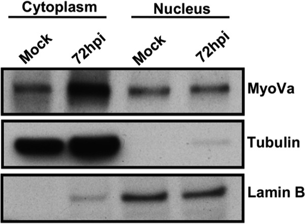
HFFs were either mock infected or infected with WT HCMV (MOI of 1). At 72 hpi, nuclear and cytoplasmic fractions were prepared and then analyzed by Western blotting to assess myosin Va (MyoVa) expression levels, with tubulin and lamin B serving as fractionation controls for the cytoplasm and nucleus, respectively.