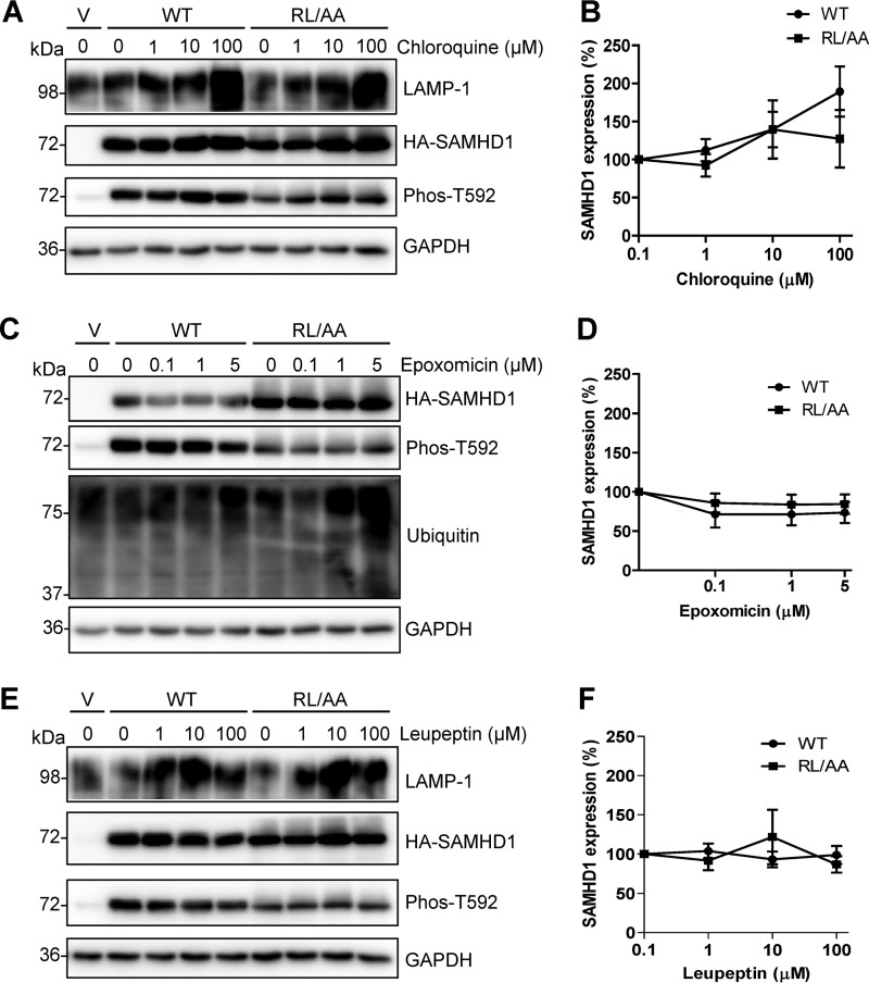
FIG 4.
SAMHD1 protein levels are not affected by inhibiting the proteasome or lysosome degradation in cells. HEK293T cells expressing WT or RL/AA SAMHD1 protein were treated with chloroquine (A), epoxomicin (C), or leupeptin (E) at the indicated concentrations for 16 h. Vector plasmid DNA-transfected cells were used as a negative control. Lysates were harvested and analyzed by immunoblotting using antibodies specific to LAMP-1 or ubiquitin, HA-tagged SAMHD1 (HA), and Phos-T592 SAMHD1. GAPDH was used as a loading control. (B, D, and F) Graphs depicting SAMHD1 were generated from densitometry analysis of WT or RL/AA SAMHD1 (HA) immunoblots in HEK293T cells treated as described for panels A, C, and E. All densitometry was normalized to the value for GAPDH. Negative controls with no inhibitor treatment were set to 100% for each WT or RL/AA experiment, and all densitometry values were calculated as a percentage of this. The graphs represent a summary of 6 independent transfection experiments.

