Figure 3.
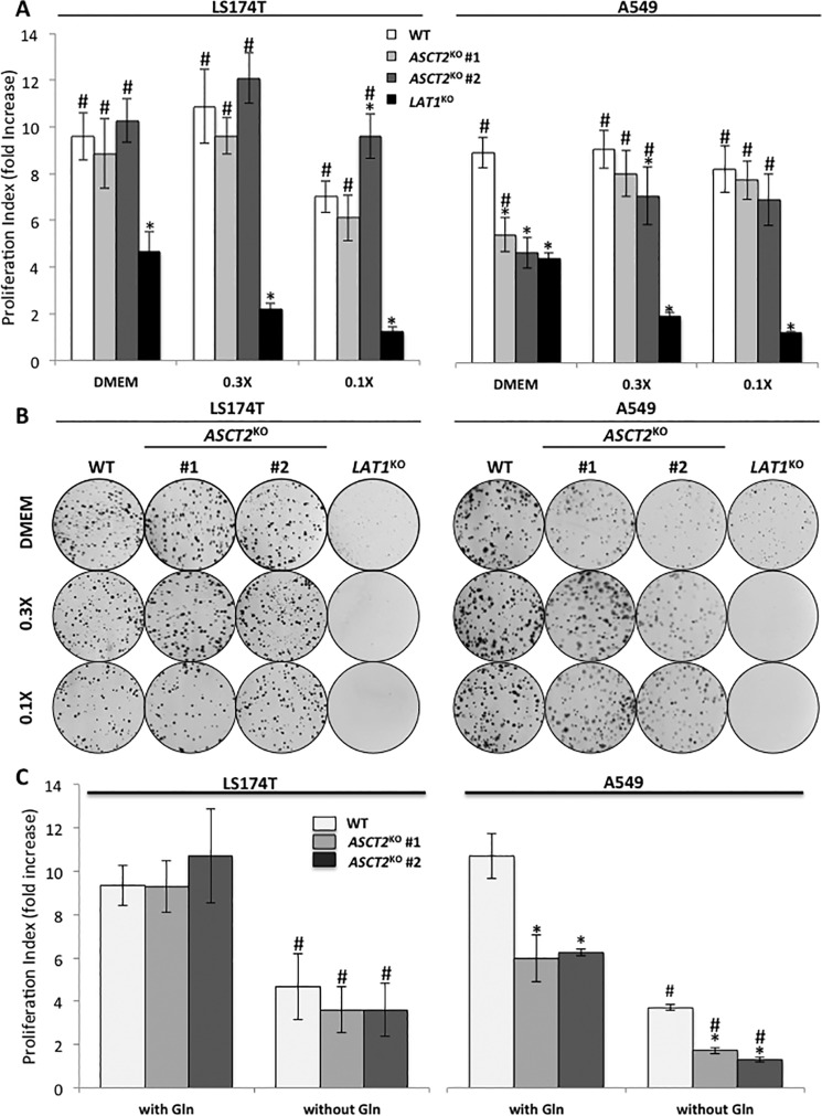
ASCT2 knock-out does not phenocopy LAT1 knock-out in vitro. A, cell proliferation of WT (white), ASCT2KO (light and dark gray, two independent clones), and LAT1KO (black) cells of LS174T and A549 cell lines. Cells were cultivated for 72 h in DMEM, 0.3×, or 0.1× media (Table S2). The media were replaced every day to maintain constant AA concentrations. Proliferation rates are presented as -fold increase (see “Experimental Procedures” for detailed description). These data represent the average of at least three independent experiments. *, significant compared with WT (ANOVA, p < 0.05), #, significant compared with LAT1KO (p < 0.05). B, clonal growth of WT, ASCT2KO (#1 and #2), and LAT1KO cells of LS174T and A549 cell lines. Cells were cultivated for 15 days in DMEM, 0.3× or 0.1× media (Table S1). The media were replaced every 2 days to maintain constant AA concentrations and colored for visualization using Giemsa. C, cell proliferation of LS174T and A549 WT (white) and ASCT2KO (light and dark gray, two independent clones) cells cultivated for 72 h in glutamine-free DMEM. These data represent the average of at least three independent experiments. *, significant compared with WT (ANOVA, p < 0.05); #, significant compared with DMEM (+) glutamine (p < 0.05).

