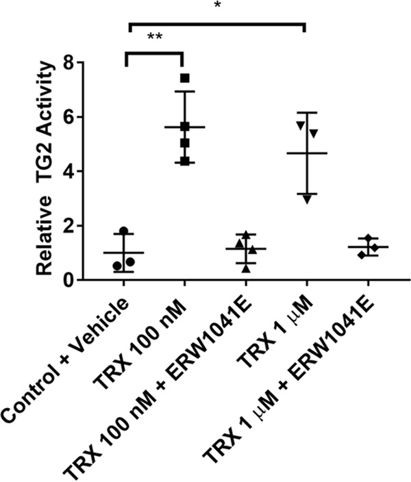
Figure 3.

Activation of extracellular TG2 by TRX in HUVECs. Cells were pre-incubated with recombinant, reduced TRX with or without 25 μm ERW1041E for 1 h. 5BP was then added, and TG2 activity was assayed for 3 h. Cells were the washed, fixed without permeabilization, and probed with streptavidin-HRP. TMB was added, and the reaction was followed continuously at 655 nm for 30 min. The data were normalized against the Control + Vehicle condition. Statistical comparisons were performed using Student's t test. A dose of 100 nm (**, p < 0.01) and 1 μm (*, p < 0.05) TRX resulted in increased TG2 activity that was reversed with TG2 inhibitor (ERW1041E). Data are presented as average ± S.D. of at least three replicates per condition.
