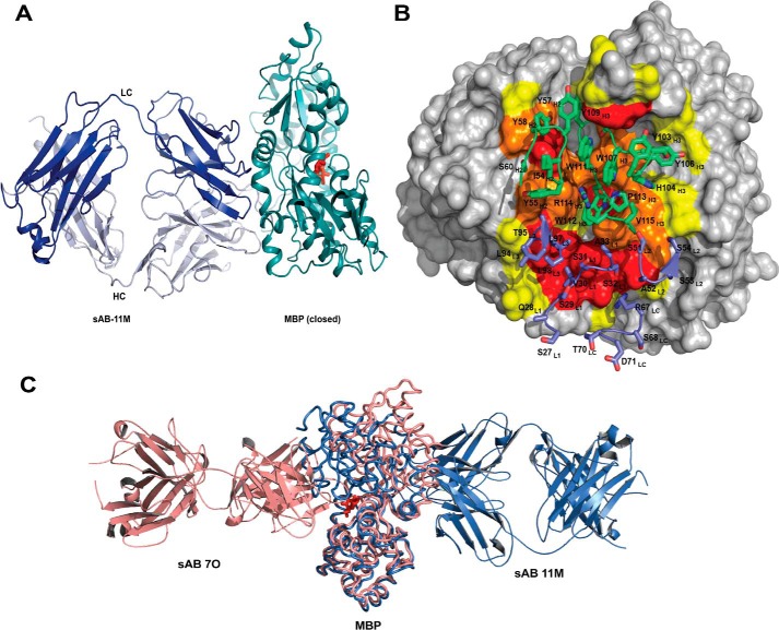
Figure 4.
Structures of closed-specific sAB-11M·MBP complex. A, sAB-11M binds to the hinge region in the maltose (red sticks)-bound conformation of MBP on the side opposite to the maltose-binding pocket. B, detailed picture of sAB-11M residues (shown in sticks) interacting with MBP. CDR-L1, -L2, and -L3 and LC scaffold residues are colored marine, and CDR-H1, -H2, and -H3 are colored green. MBP is color-coded as in Fig. 3B. C, superposition of structures of sAB-7O-MBP structure (salmon) with sAB-11M-MBP (slate) shows that sAB-7O and sAB-11M bind on opposite sides of MBP in open and closed states, respectively.

