Figure 2.

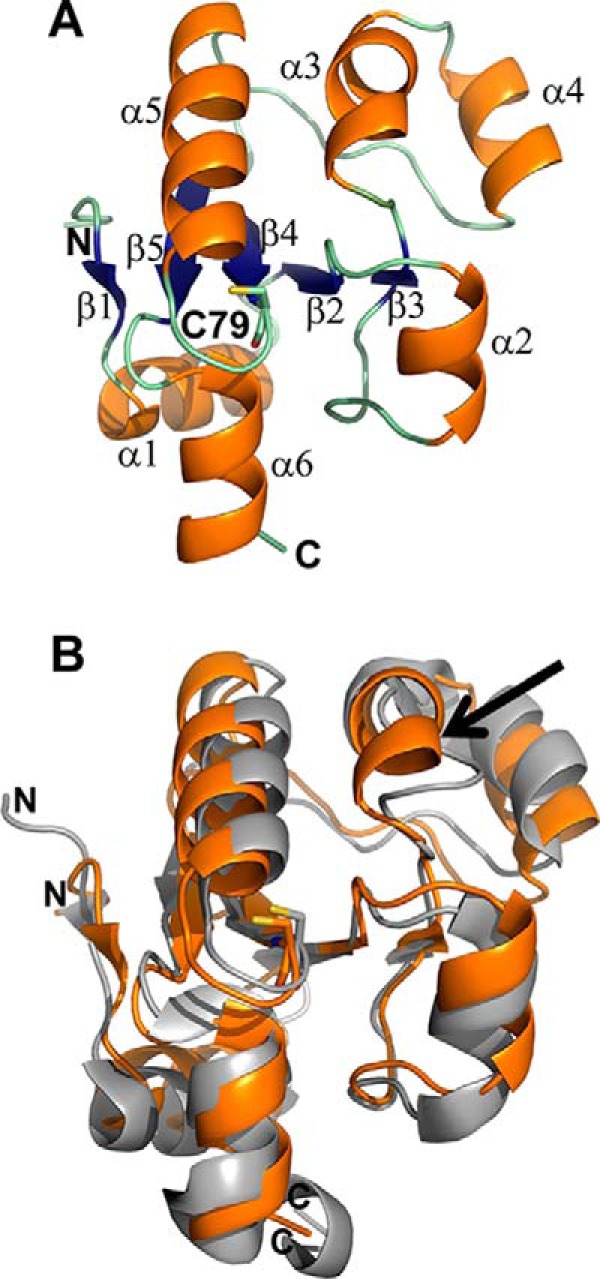
Crystal structure of TSTD1. The X-ray crystal structure of TSTD1 was solved at 1.04 Å resolution by SAD phasing with SeMet. A, the structure of the TSTD1 monomer consists of a five-stranded parallel β-sheet core (blue) surrounded by six α-helices (orange; α1–6). The active-site cysteine, Cys-79, is shown in a stick representation. B, comparison of TSTD1 with RDL1. Structural overlay of TSTD1 (orange) with the S. cerevisiae homolog RDL1 (PDB code 3D1P; gray). The active-site cysteine residues are shown in stick representation. The presence of an additional α-helix, α3, in TSTD1 is indicated by the arrow.
