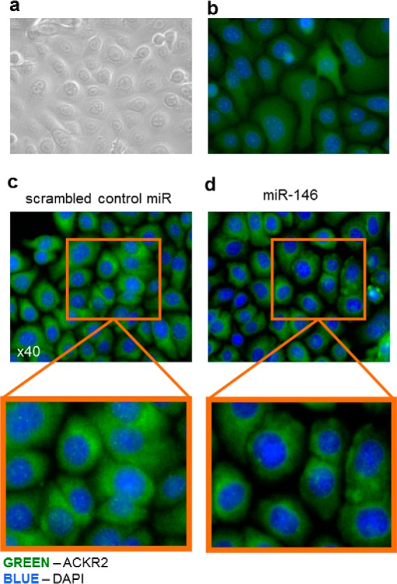
Figure 4.

Transfection of KCs with miR-146b reduced cytoplasmic ACKR2 protein distribution. a, representative bright-field image of KCs grown as a confluent monolayer. b, representative immunofluorescence of KCs grown as confluent monolayers and stained with isotype control antibody. c and d, representative immunofluorescence microscopy images of confluent monolayers of KCs 48 h after transfection with scrambled miR control (c) or miR-146b (d).
