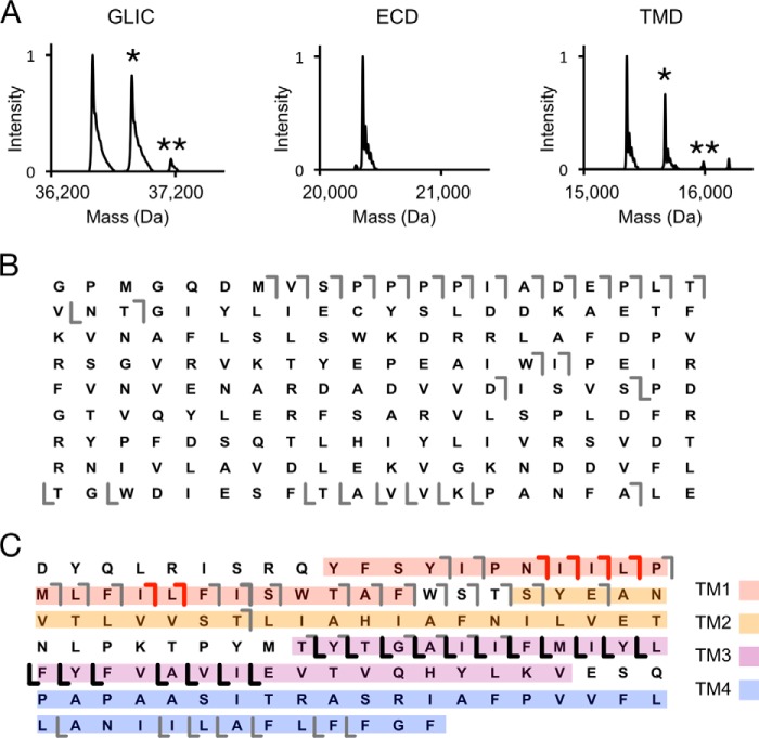
Figure 2.
AspN middle-down analysis of GLIC photolabeled with 5α-6-AziP. A, deconvoluted spectra of GLIC photolabeled with 300 μm 5α-6-AziP showing intact GLIC and the ECD and TMD after AspN digestion. B, HCD fragment ion assignments of the unlabeled ECD peptide shown in A. The gray lines represent b- and y-ions that do not contain 5α-6-AziP. C, HCD fragment ion assignments of the singly labeled TMD species in A. The red and black lines represent b-ions and y-ions, respectively, that contain 5α-6-AziP.

