Figure 5.
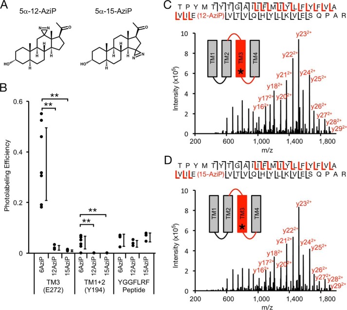
MS analysis of GLIC photolabeled with 5α-12-AziP and 5α-15-AziP. A, structure of the photolabeling reagents 5α-12-AziP and 5α-15-AziP. B, photolabeling efficiency of 5α-6-AziP, 5α-12-AziP, and 5α-15-AziP for TM3 and TM1 + 2 of WT GLIC, and the peptide YGGFLRF (n = 7 for 5α-6-AziP, n = 3 for 5α-12-AziP and 5α-15-AziP). ±S.D., **, p < 0.01. C, HCD MS2 spectrum of TM3 peptide labeled with 5α-12-AziP from GLIC WT. Red and black fragment ions do and do not contain 5α-12-AziP, respectively. D, same as C for 5α-15-AziP.

