Figure 2.
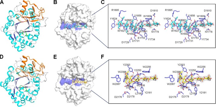
Crystal structures of OfChtII-C1 and OfChtII-C2 in free form and in complex with oligosaccharide. A and D, cartoon representation of OfChtII-C1 (A) and OfChtII-C2 (D). The structure consists of two domains: a core (β/α)8 TIM-barrel fold (cyan, α-helices; blue, β-strands) and an insertion domain (orange). The catalytic residues are shown as sticks with green carbon atoms. B and E, surface representations of OfChtII-C1 complexed with (GlcNAc)7 (B) and OfChtII-C2 E2180L complexed with (GlcNAc)5 (E). The ligands are shown as sticks with cyan and yellow carbon atoms, respectively. The aromatic residues that stack with the sugar rings are shown in blue. C and F, stereoview of the substrate-binding cleft with details of the interactions between (GlcNAc)7 and OfChtII-C1 (C) and between (GlcNAc)5 and OfChtII-C2 E2180L (F). The ligands are represented as sticks with cyan and yellow carbon atoms, respectively. The 2Fo − Fc electron-density map around the ligand is contoured at the 1.0 Å level. The catalytic residues and the amino acids that interact with the ligand are labeled and shown as sticks with blue carbon atoms. The numbers indicate the subsite to which the sugar is bound. Hydrogen bonds are drawn as dashed lines.

