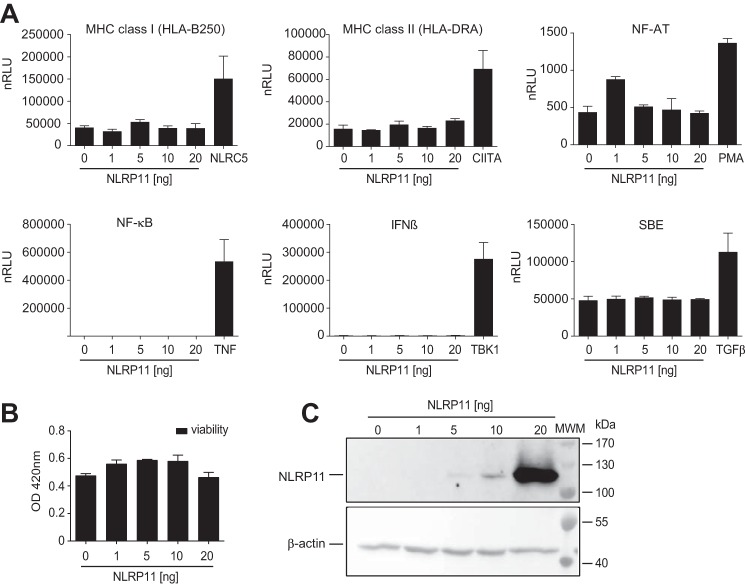
Figure 3.
Overexpression of NLRP11 does not trigger pro-inflammatory pathways. A, results from reporter assays with HEK293T cells transfected with reporter plasmids encoding the luciferase gene under the control of different promoters and co-transfected with either 0, 1, 5, 10, or 20 ng of pNLRP11.MYC plasmid. As a control, cells were transfected or stimulated as indicated (plasmid transfection, 5 ng of NLRC5, 5 ng of CIITA, or 20 ng of TBK1 plasmid; treatment, 20 ng/ml TNF, 100 ng/ml TGFβ, or 100 nm PMA). Relative light units normalized to β-galactosidase expression (nRLU) are shown. Error bars represent standard deviation of the mean of representative experiments conducted in triplicates of three independent experiments. B, XTT cell viability assay 16 h after overexpression of NLRP11 in HEK293T cells. Measurement of the optical density (OD) at 450 nm (mean and standard deviation of two independent experiments). C, Western blot from protein lysates from HEK293T cells probed with anti-myc antibody and β-actin as a loading control. **, p < 0.005; ***, p < 0.0005. MWM, molecular weight marker.

