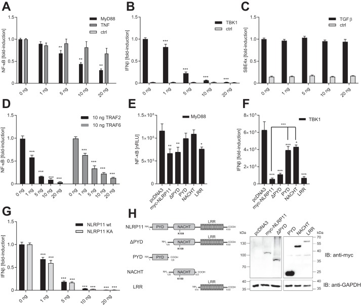
Figure 4.
NLRP11 overexpression negatively affects innate immune signaling pathways. Gene reporter assays with HEK293T cells transfected with the indicated reporter and NLRP11 expression plasmids. A–D, effect of increasing expression of NLRP11 on inflammatory pathways. A, cells were activated by transfection of 20 ng of MyD88 plasmid or incubation with 20 ng/ml TNF or not treated (control (ctrl)). NF-κB-promoter activity is shown. B, cells were activated by transfection of 20 ng of TBK1 plasmid or empty plasmid (ctrl). IFNβ promoter activity is shown. C, cells were activated with 100 ng/ml TGFβ or not treated (ctrl). Smad-binding element promoter activity is shown. D, cells were activated by transfection of 10 ng of TRAF2 or TRAF6 plasmid. NF-κB promoter activity is shown. E, cells were transfected with the indicated plasmids and activated by transfection of 20 ng of MyD88 plasmid. NF-κB promoter activity is shown. nRLU, normalized relative light units. F, cells were transfected with the indicated plasmids and activated by transfection of 20 ng of TBK1 plasmid or empty plasmid (no TBK1). IFNβ promoter activity is shown. G, effect of the Walker A mutant of NLRP11. Cells were activated by transfection of 20 ng of TBK1 plasmid or empty plasmid (ctrl). IFNβ promoter activity is shown. H, schematic and expression analysis by immunoblot (IB) of the constructs of NLRP11 used in E and F. -Fold reduction relative to control cells transfected with 0 ng of NLRP11 set as 1 (A–D) or normalized relative light units are shown (E–G). Error bars represent standard deviation of the mean of three experiments, each conducted in triplicate. *, p < 0.05; **, p < 0.005; ***, p < 0.0005; n.s., p > 0.99.

