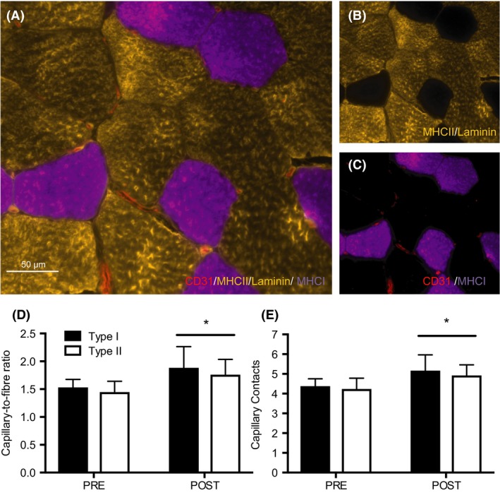
Figure 1.

Capillary expression in type I and II muscle fibers after 6 weeks of HIIT. Representative image of a CD31/MHCII/laminin/MHCI stain (A). Co‐expression of MHCII/laminin (gold) (B), and CD31/MHCI (red/purple) (C). Capillary‐to‐fiber ratio (D) and capillary contacts (E) before (PRE) and after (POST) training in type I and type II fibers. Data are presented as means ± SD. *P < 0.05, main effect of time. HIIT, high‐intensity interval training.
