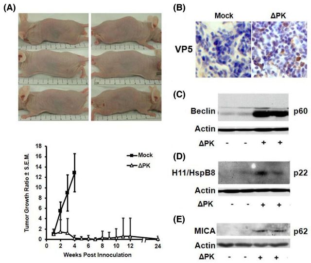
Figure 4.
ΔPK inhibits the growth of LM melanoma xenografts. (A) LM melanoma cells (107) were implanted subcutaneously into both flanks of Balb/c nude mice and given four intratumoral injections of ΔPK (n = 4; 106 pfu) or growth medium (n = 4; mock) at weekly intervals beginning on day 7, when the tumors were palpable. Tumor volume was monitored for 5 months after the last ΔPK injection. The difference between mock and ΔPK treatment became statistically significant on day 14 (p < 0.001 by two-way ANOVA) and remained significant to the end of the study. Three ΔPK-treated mice showing complete tumor eradication were photographed at day 35. Data are expressed as tumor growth ratio, calculated by dividing each tumor volume measured over time by the initial tumor volume on day 7. (B) A2058 xenografts mock or ΔPK infected were collected at 7 days after the last ΔPK injection and stained with antibody to the major virus capsid protein VP5 by immunohistochemistry and counterstained with Mayer's Hematoxylin. Duplicates of the A2058 xenografts were immunoblotted with antibodies to Beclin-1 (C), H11/HspB8 (D) or MICA (E), and then stripped and re-probed with antibody to actin. Each lane represents a different tumor. Representatives are shown for each antibody. IACUC approved protocol #0513003.

