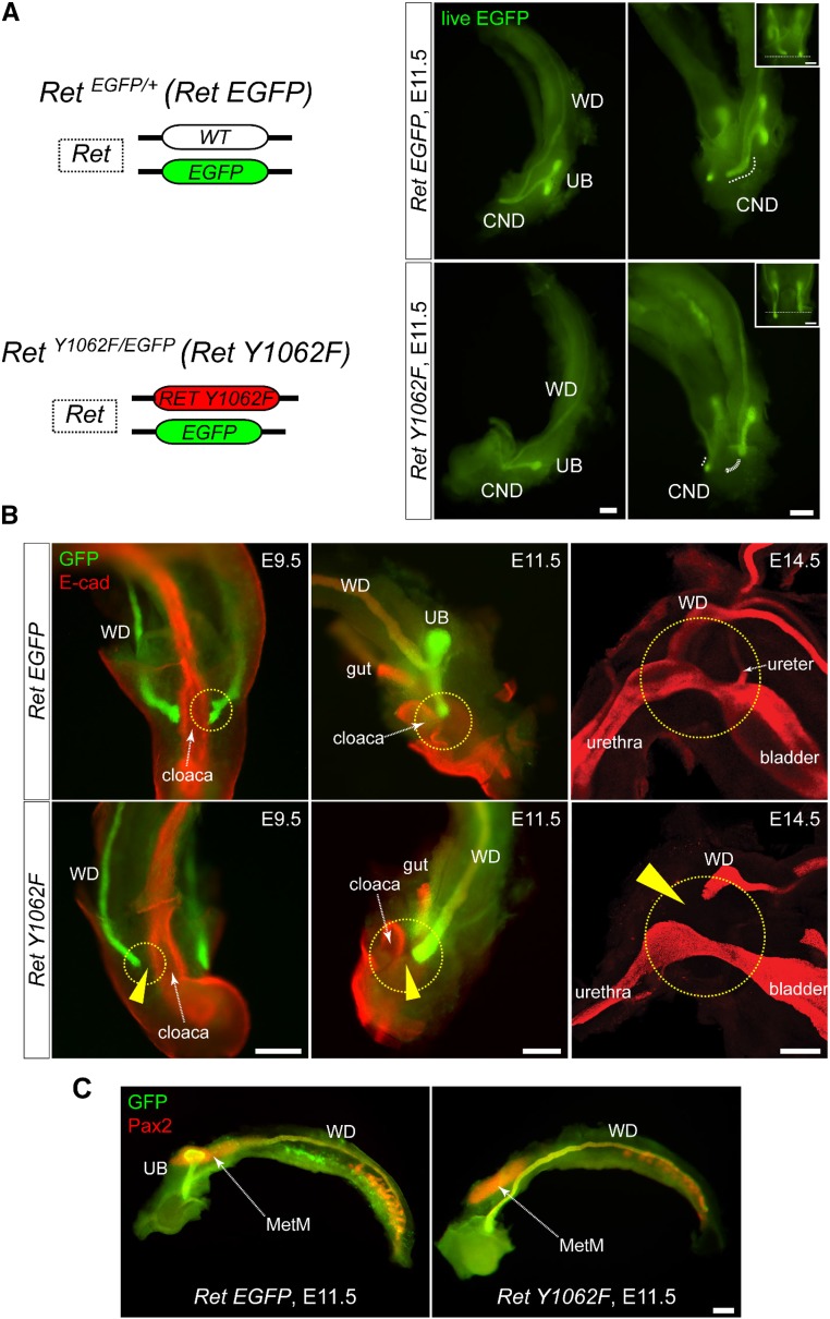
Figure 1.
WDs of RetY1062F mutant mice fail to reach cloaca. (A) Left: Simplified illustration of modified Ret locus denoting RetEGFP reporter and RetY1062F mutant alleles. Right: Live EGFP (green) images of whole urinary tract of RetEGFP (RetEGFP/+) control and RetY1062F (RetY1062F/EGFP) embryos at embryonic day 11.5. Although the CND and T-shaped UB are normally developed in control (top), the UB is absent or rudimentary with no branching in RetY1062F mice (bottom, see also Supplemental Movie 1). WD length was asymmetric in 57% of the mutants (13 out of 23 embryos, inset) denoting defective elongation. (B) Whole-mount immunofluorescence images with GFP (green, WD) and E-cad (red, cloaca or bladder) antibodies reveal that the mutant WDs fail to reach cloaca or bladder during development. Images for embryonic days 9.5 and 11.5 are whole-mount images acquired by fluorescence stereomicroscope, and 14.5 images are three-dimensionally reconstructed confocal microscopy images. Arrowheads show the gap between the WD tip and cloaca or bladder. (C) Whole-mount immunofluorescence images with GFP (green, WD and UB) and Pax2 (red, WD, UB, and MetM) antibodies show normally developed MetM in RetY1062F mutant urinary tract, indicating that the WD defects seen in this mutant are autonomous. Scale bar, 200 μm. MetM; metanephric mesenchyme.

