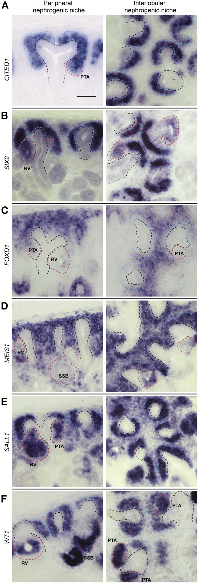
Figure 1.
In situ hybridization labeling for nephron compartment marker genes. (A-F) show expression for genes as indicated on fields. Left-hand and right column fields display in situ hybridization labeling of cryo-sectioned human week 14–15 kidneys. Sections show peripheral nephrogenic niches and interlobular nephrogenic niches (left and right, respectively). Red, blue, and black dashed lines indicate nascent nephrons, cap mesenchyme, and ureteric bud epithelium, respectively. PTA, pretubular aggregate; RV, renal vesicle; SSB, S-shaped body. Scale bar 50 μm.

