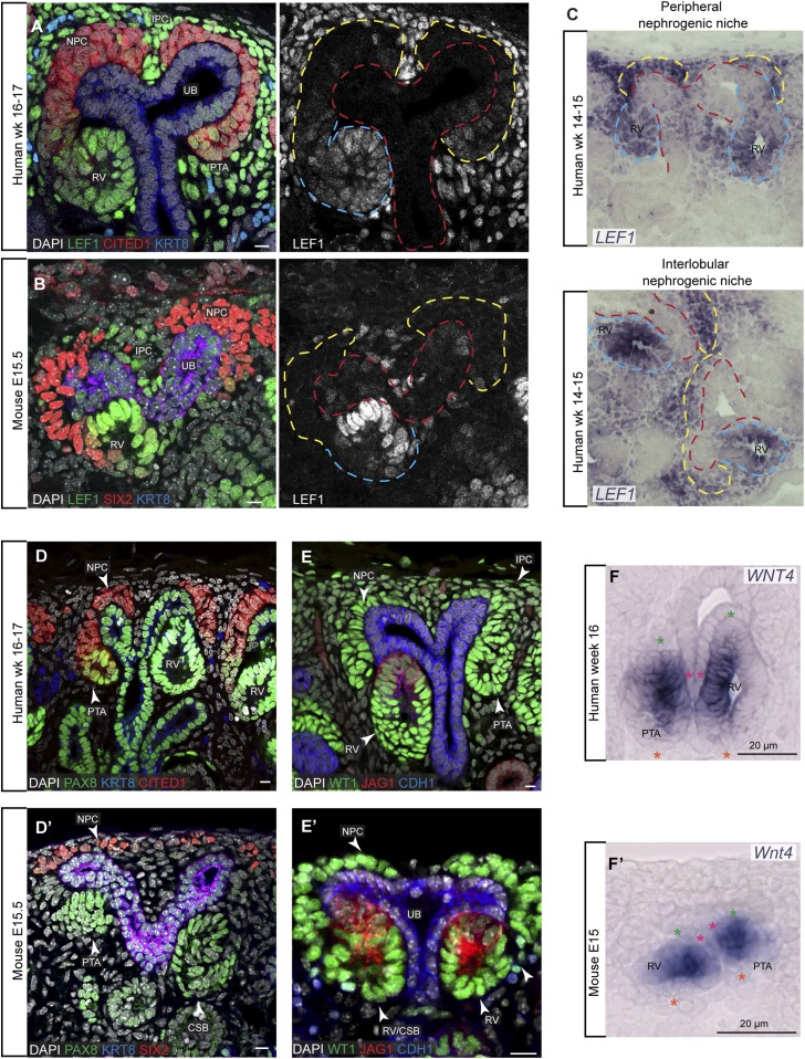
Figure 1.
Nephron progenitor induction in human and mouse nephrogenic niches. (A–F) and (D’–F’) Immunofluorescent stains and in situ hybridization on human and mouse kidneys, respectively. Ages and stains as specified on fields. Yellow, red, and cyan dashed lines indicate cap mesenchyme, ureteric bud, and nephrons, respectively. Stars in (F) and (F’) indicate the nephron axes: green, distal; orange, proximal; magenta start indicates ureteric bud. Scale bars on immunofluorescence data indicate 10 µm. For single-channel views see Supplemental Figures 1 and 3. CSB, comma-shaped body; IPC, interstitial progenitor cells; UB, ureteric bud.

