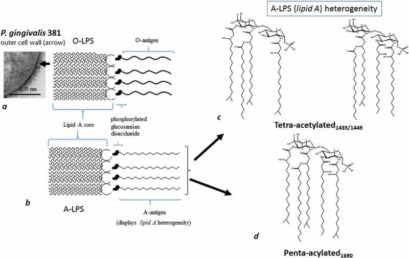
Figure 1.

Schematically shows two main forms of LPS (O-LPS and A-LPS) and their position on the outer cell wall (arrow). (a) Section of P. gingivalis (FDC 381) demonstrated by transmission electron microscopy. Thick arrow points to lipid A with fatty acid esters attached to phosphorylated glucosamine disaccharides and the O-antigen. The latter is O-LPS (with O-antigen tetrasaccharide repeating units); (b) A-LPS (with surface anionic polysaccharide [APS] repeating units). There are two lipid As in A-LPS as shown in (c) and (d). (c) A tetra-acylated form with two different molecular weights, hence two structures. (d) the penta-acylated form.
