Figure 1.
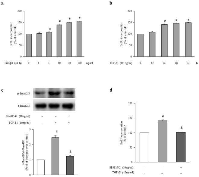
TGF-β1 stimulates ASMCs proliferation via activation of Smad2/3. (a) ASMCs were stimulated with different concentrations of TGF-β1 ranging from 0 to 100 ng/ml for 24 h, the rate of BrdU incorporation in cells was determined by BrdU ELISA assay Kit (n = 4 per group). (b) Cells were exposed to 10 ng/ml TGF-β1 for the indicated times, BrdU incorporation in cells was measured (n = 4 per group). (c) ASMCs were treated with SB431542 (10 μM) for 1 h before stimulation with TGF-β1 (10 ng/ml) for 1 h, the phosphorylation of Smad2/3 was determined by immunoblotting (n = 4 per group). The full-length blots of Fig. 1c are presented in Supplementary Fig. S1. (d) ASMCs were treated with SB431542 (10 μM) for 1 h and then stimulated with TGF-β1 (10 ng/ml) for 24 h, BrdU incorporation in cells was measured (n = 4 per group). *P < 0.05 versus control. #P < 0.01 versus control. &P < 0.01 versus TGF-β1-treated cells.

