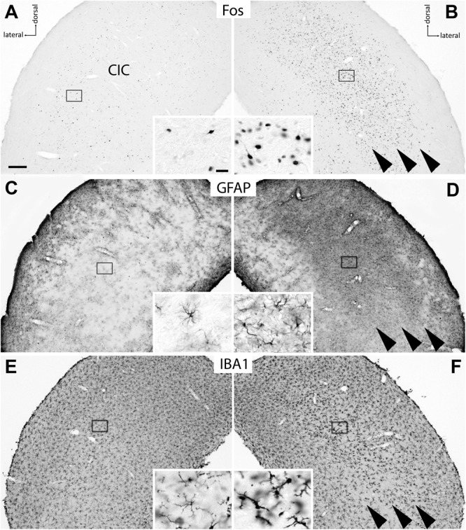
FIGURE 2.

Effect of 1 d EIS on cells in the CIC of deafened rats. (A,B) Fos was expressed in the CIC contralateral to the simulated cochlea (B) in a population of neurons that extends beyond tonotopic boundaries known from nh animals (arrowheads). Ipsilaterally (A), only few and scattered Fos-positive neurons were present. (C,D) In a nearby section stained for GFAP, a massive growth of astrocytes was noted taking place regionally specific in the contralateral CIC where Fos-positive neurons were abundant (D, arrowheads); no such changes occurred ipsilaterally (C). (E,F) In another nearby section, microglia identified by IBA1 immunoreactivity showed a modified morphology indicating their activation in just the same region of the contralateral CIC (F, arrowheads); no such changes occurred ipsilaterally (E). Frames show positions of insets. Scale bar for major panels: 200 μm; scale bar for insets: 20 μm.
