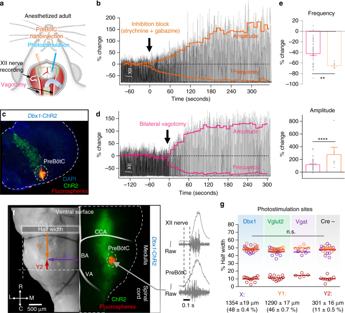
Fig. 6.
Effects of preBötC sensory feedback inhibition on hypoglossal motor output in vivo. a Schematic of the surgical approach to access the ventral brainstem for bilateral preBötC nanoinjections, photostimulation, and vagotomy. b Representative trace of integrated hypoglossal (XII) nerve activity during bilateral injection of strychnine and gabazine (150 nl, 250 µM each) with frequency and amplitude overlaid (10 s bins). c Example transverse hemisection showing injection site marked by flourospheres localized to the preBötC in a Dbx1-ChR2 mouse. d Representative trace of XII activity during bilateral vagotomy with frequency and amplitude overlaid (10 s bins). e Quantified changes in frequency and amplitude ~5 min following vagotomy (n = 47) and blockade of preBötC fast synaptic inhibition (n = 6; unpaired two-tailed t-tests with Welch’s correction; **p < 0.01, ****p < 0.0001). f Bright field image (left) and Dbx1-ChR2 fluorescence (middle) of the ventral medulla showing the location of preBötC photostimulation relative to the basilar artery (BA), caudal cerebellar artery (CCA), and intersection of the vertebral arteries (VA). Example extracellular recording demonstrating pre-inspiratory activity relative to XII nerve activity at photostimulation sites (right). g Quantified coordinates of injected fluorospheres (bottom) demonstrate that photostimulation sites were consistent across experimental groups (Dbx1, n = 14; Vglut2, n = 14; Vgat, n = 5; Cre-, n = 10; two-way ANOVA and Bonferonni’s post hoc test; n.s., not significant, p > 0.05)

