Figure 1.
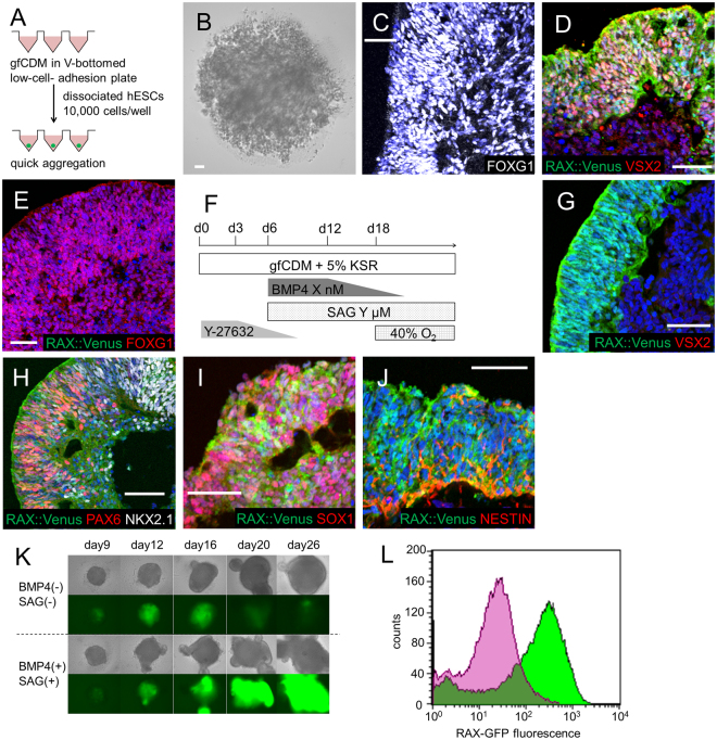
Differentiation into RAX+ hypothalamic progenitors. (A) Schematic of 3D culture (SFEBq) start conditions. (B) Failure of aggregation in SFEBq with gfCDM without KSR. (C) FOXG1 (white) expression in day 20 aggregates cultured with gfCDM containing KSR. FOXG1 is a telencephalic marker. (D) Induction of neural retina in day 30 aggregates in SFEBq with gfCDM + KSR and with BMP4 treatment. RAX::Venus (green), VSX2 (red). (E) Increased FOXG1 expression in day 20 aggregates treated with SAG in gfCDM + KSR. RAX::Venus (green), FOXG1 (red). (F) Culture protocol for induction of hypothalamic progenitors. (G–J) Immunostaining of day 30 aggregates with both BMP4 and SAG treatment in gfCDM + KSR. (G) RAX::Venus (green)+, VSX2 (red) −indicated hypothalamic progenitors. (H) Mixed pattern of dorsal (PAX6+) and ventral (NKX2.1+) hypothalamic precursors. RAX::Venus (green), PAX6 (red), NKX2.1 (white). (I,J) Expression of neural ectoderm markers. RAX::Venus (green), SOX1 (I, red) and NESTIN (J, red). (K) Time courses of RAX::Venus expression. Upper panels: bright field view, lower panels: fluorescence views of RAX::Venus+ cells. (L) FACS-analysis of day 20 aggregates. Over 80% of cells were induced to RAX+ hypothalamic progenitors. Green: BMP4 (+)/SAG (+), Purple: BMP4 (−)/SAG (−). For all relevant panels, nuclear counter staining: DAPI (blue), scale bar: 50 μM.

