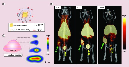
Figure 6. . PET/CT imaging of 64Cu-DOTA-PEG-Au nanocages in an EMT-6 tumor mouse model.
(A) Schematic illustration of the surface-modified Au nanocages; (B) PET/CT images of 30 nm 64Cu-DOTA-PEG-AuNCs in a mouse bearing an EMT-6 tumor at 1, 4 and 24 h post injection (3.7 MBq injection per mouse). T: tumor; B: bladder. (C) PET/CT images recorded from different cross-sections of an EMT-6 tumor, showing the intratumoral distribution of Au nanocages [53].
Reproduced with permission from [53].

