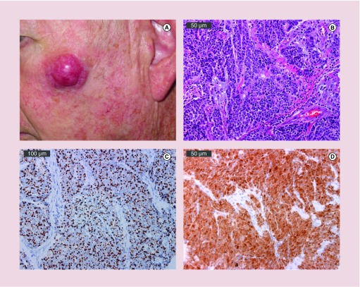
Figure 1. . Clinical and pathologic presentation of Merkel cell carcinoma.
(A) A 2.5 cm primary MCC on sun exposed skin of the left cheek. (B) Hematoxylin & eosin magnification of MCPyV-positive MCC tumor. Bar indicates 50 μm. (C) Cytokeratin-20 immunohistochemical staining of an MCPyV-positive MCC demonstrates characteristic perinuclear dot-like expression. Bar indicates 100 μm. (D) Viral oncoprotein expression limited to tumor (not adjacent stroma). MCPyV LT antigen expression detected using CM2B4 antibody. Bar indicates 50 μm. Photos courtesy of Chris Lewis.

