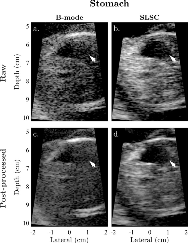
Figure 2.

Example matched (a) raw B-mode, (b) raw SLSC, (c) post-processed B-mode, and (d) post-processed SLSC images of the fetal stomach shown with sonographer-optimized parameters for dynamic range and short-lag. Arrows indicate the lateral boundary of the stomach, which is significantly degraded by reverberation clutter in B-mode. SLSC imaging appears to reduce the appearance of this clutter, resulting in improved conspicuity and definition of the stomach and its boundaries.
