Fig. 1.

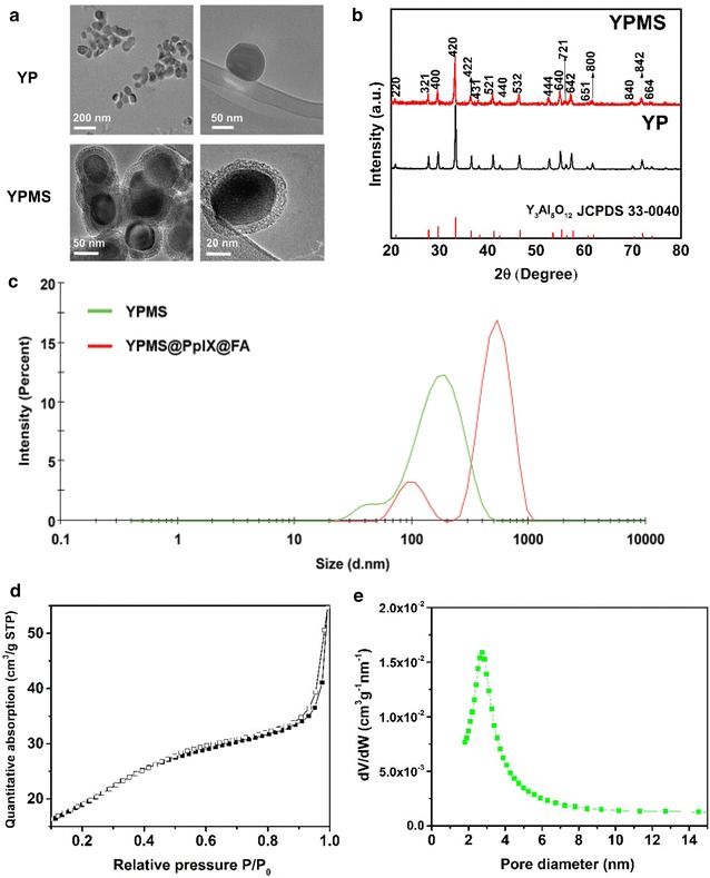
Morphological characterization YPMS nanoparticles. a Transmission electron microscopy images of a bare YP and YP nanoparticles covered with mesoporous silica (YPMS). b X-ray diffraction pattern of YP bare or covered with mesoporous silica, YPMS. Values correspond to hkl coordinate of the YAG cubic crystal. c Hydrodynamic diameters of YPMS and YPMS@PpIX@FA in water using dynamic light scattering. d Nitrogen adsorption–desorption isotherms of YPMS nanoparticles. Filled square adsorption, empty square desorption. e Pore size distribution of the mesoporous layer of YPMS nanoparticles
