Figure 7.

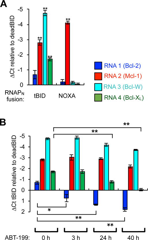
Detection of 1×4 Bcl-2 family PPIs simultaneously in mammalian cells by RT-qPCR analysis of the unique RNA outputs. (A) HEK293T cells were cotransfected with the plasmids shown in Figure 6A with the fusions indicated, grown for 40 h, lysed, and then total RNA was isolated and quantified by RT-qPCR. Separate PCR primers were used for each of the four unique RNA outputs to measure split RNAP assembly with each target. The data displayed is the delta-Ct value in comparison to cells transfected with the RNAPN-deadBID “negative control”. Therefore, a more negative value indicates more of a particular RNA is generated, and therefore more of a given interaction was present. (B) HEK293T cells were cotransfected with the plasmids shown in (Figure 6A) with the tBID-RNAPN fusion, grown for 40 h with 0.5 μM ABT-199 added at different time points, lysed, and then total RNA was isolated and quantified by RT-qPCR as described in (A). The data displayed is the delta-Ct value in comparison to cells transfected with the RNAPN-deadBID “negative control”. Error bars are ± s.e.m., n = 4. Student’s t-test; *P < 0.05, **P < 0.0005.
