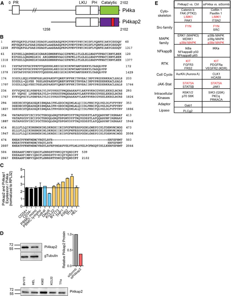
Figure 7. The Human PI4KAP2 Gene Yields a Protein Product and Has Higher mRNA Expression Relative to PI4KA in Erythro- and Myelo-Leukemia Cell Lines.

(A) Schematic comparing PI4KA and PI4KAP2 proteins. PI4KAP2 lacks the N-terminal domain and has a deletion in the kinase domain (red). PR, proline-rich domain; LKU, lipid kinase unique domain; PH, plekstrin homology domain.
(B) Alignment of PI4KA and PI4KAP2 amino acid sequences shows major homology starting at amino acid 1,258 of PI4KA, except for missing amino acids in the kinase domain of PI4KAP2.
(C) Ratio of mRNA expression of PI4KAP2 and PI4KAP1 compared with PI4KA in a panel of normal cord and peripheral blood cells (black), human lymphoid leukemia (blue), and myeloid leukemia (orange) cell lines.
(D) Top: Validation of an antibody probing cells subjected to siRNA targeting PI4KAP2 and quantification. Bottom: Western blot showing endogenous PI4KAP2 in myeloid leukemia cell lines.
(E) Antibody array summary of proteins that change when lysates from cells overexpressing PI4KAP2 were compared with vector C (left) and of proteins that change when lysates from cells knocked down for PI4KA were compared with C cells (right). Proteins in common between the two comparisons are highlighted in red. See also Figures S6 and S7.
