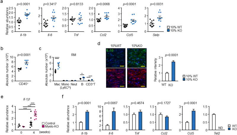
Figure 3. Effect of Tet2-deficient hematopoietic cells on the expression of pro-inflammatory cytokines and chemokines in the remodeling heart tissue.
a. Analysis of transcript expression in the non-infarcted marginal zone obtained from 10% KO-BMT mice (n=10) and 10% WT-BMT (n=11) mice. Gene expression was analyzed by qPCR analysis. Statistical significance was evaluated by two-tailed unpaired Student’s t tests with Welch’s Correction when variance was unequal or by Mann Whitney U tests for data which failed to pass the Shapiro-Wilk normality test. b. Flow cytometry analysis of cardiac remote area from 10% KO-BMT mice (n=7) and 10% WT-BMT (n=7) mice to show the absolute number of total CD45+ immune cells are increased in the myocardial tissue from 10% KO-BMT mice. Data is expressed as number of cells per 100 mg wet weight. Statistical analysis was evaluated by two-tailed unpaired Student’s t test. c. Flow cytometry analysis of cardiac remote area from 10% KO-BMT mice (n=7) and 10% WT-BMT (n=7) mice to show the absolute number of each immune cell populations. Statistical significance of difference was evaluated by multiple t tests. d. IL-1β immunofluorescence staining in Mac3-positive macrophage-enriched marginal zone of 10% KO-BMT (n=5) mice and 10% WT-BMT mice (n=5) showing IL-1β signal is higher in 10% KO-BMT mice. Scale bars = 20 µm. Images were quantified for integrated fluorescence intensity with Image J software. Statistical analysis was performed by two-tailed unpaired Student’s t tests. e. Remote area samples were obtained from conditional myeloid-specific KO mice and control mice, and gene expression was analyzed by qPCR at the indicated time points (n=3 for sham and n = 10 at 4 weeks after LAD ligation, per genotype). Statistical significance was evaluated by two-way ANOVA with Tukey’s multiple comparison tests. f. Bone marrow-derived macrophages 2 days after in vitro differentiation obtained from Tet2-null mice (n=6) and wild type (n=7) were obtained in vitro and gene expression was analyzed by qPCR analysis. Statistical significance of difference was evaluated by two-tailed unpaired Student’s t tests with Welch’s Correction when variance was unequal or by Mann Whitney U tests for data which failed to pass the Shapiro-Wilk normality test. *p<0.05, **p<0.01, ****p<0.0001. WT: wild type, KO: knockout, BMT: bone marrow transfer, LAD: left anterior ascending artery, qPCR: quantitative polymerase chain reaction, RM: remote area, Mac: macrophages, Mono: monocytes, Neut: neutrophils, B: B cells, T: T cells, ND: not detected.

