
Fig. 6.
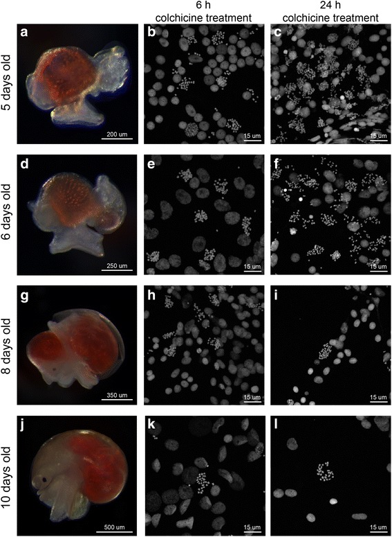
Chromosome spreads for different embryonic stages of P. canaliculata. The duration of colchicine treatment on embryos resulted in different densities of mitotic chromosomes for different embryonic stages. a, d, g, j P. canaliculata embryos 5, 6, 8, and 10 dpf, respectively, after removal of egg shell and perivitelline fluid. b, e, h, k Chromosome spreads obtained from embryos at indicated developmental stages incubated in colchicine for 6 h. c, f, i, l Chromosome spreads obtained from embryos at indicated developmental stages incubated in colchicine for 24 h