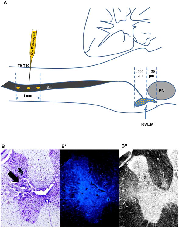
Figure 1. Fluorogold injections at T9-T10 of the spinal cord.

A) Schematic representation of Fluorogold injection (1mm from the dorsal surface - gold ovals) into the intermediolateral cell column (IML) of the spinal cord. Fluorogold (5%) was injected using a glass micropipette at the level of T9-T10, where neurons from the RVLM (gold filled circles) send their projections. Fluorogold labeled neurons were found throughout the rostro-caudal extent of the RVLM. Fewer cells were captured from 150μm rostral to caudal pole of Facial Nucleus (FN) compared to the region of the RVLM that lie 500μm caudal to the caudal pole of the FN. Diagram modified from Dampney, 1994. B) Representative Cresyl violet stained section of the spinal cord showing the injection site (black arrow) at the level of T9. B′) Fluorescent section adjacent to section in Figure B, showing the fluorescence intensity in the injection site (white arrow) at the level of T9. B”) Bright field image of the same section shown in B′ demonstrating the changes in tissue architecture at the level of injection site (black arrow). Scale bar, 100 μm. cc=central canal; DH=dorsal horn; IML=intermediolateral cell column; VH=ventral horn.
