
Figure 4. Bulbospinal catecholaminergic neuron from the RVLM of a physically active rat.
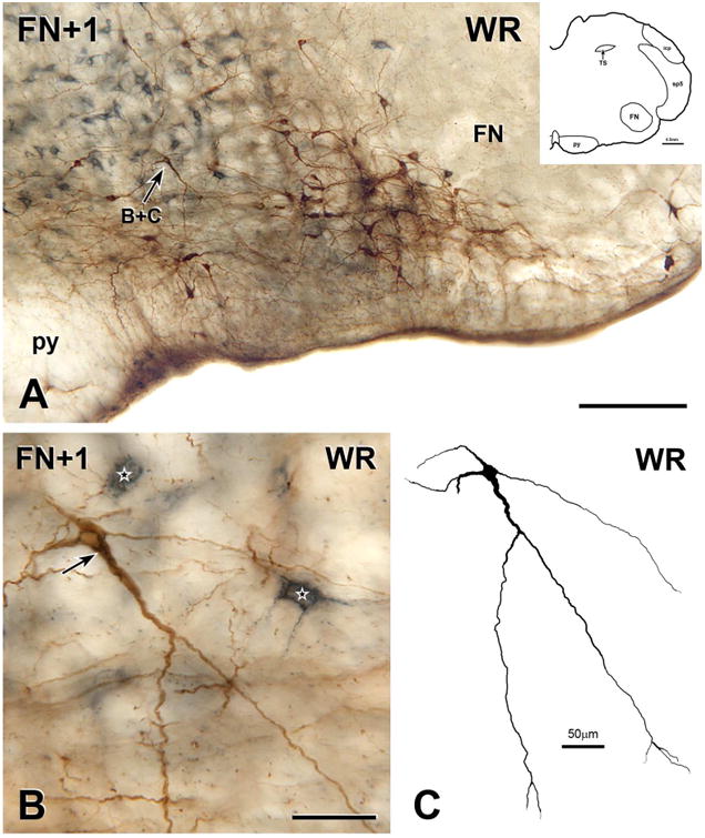
A, Montage of 5 photomicrographs showing a section through the RVLM located 150 μm rostral to the caudal pole of the facial nucleus (FN+1) from the medulla of a rat that ran voluntarily on an in-cage running wheel for 11 weeks. The section contains many neurons that are immunoreactive for both CTB (blue-grey) and TH (brown), one of which is indicated by arrow B+C. Scale bar, 250 μm. Inset, Drawing of FN+1 showing architectonic landmarks. B, Montage of 11 photomicrographs showing the double-labelled neuron indicated by arrow B+C in A. The neuron contains a network of blue-grey staining (arrow) in its cell body and in a thick proximal dendrite, indicating that the neuron has retrogradely transported CTB from the spinal cord. Two other retrogradely labelled neuron (stars) that lacks TH-immunoreactivity lie near to the bulbospinal TH neuron. Scale bar, 50 μm. C, Digital reconstruction of the neuron in B. Scale bar, 50 μm.