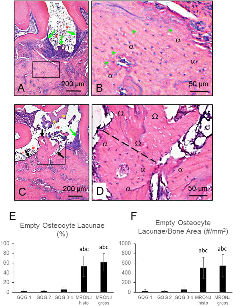
Fig. 6.
Histologic Features of FILP and MRONJ lesions and osteocyte lacunae data in ZOL-treated rats. Five-µm sections stained with H&E. B and D are magnified fields of the rectangles demarcated in A and C, respectively. (A & B) FILP lesion with GQG3 with vital alveolar bone characterized by basophilic osteocyte nuclei within lacunae (α) and only a few scattered, empty lacunae (green arrowheads [B]); substantial hyperplasia of the gingival epithelium (green arrows [A]), inflammatory cell infiltration of the lamina propria (†), disruption of the periodontal ligament (PDL), apical migration of the junctional epithelium, and alveolar bone crest resorption. (C and D) MRONJ lesion with bone exposed to the oral cavity (black arrows) with adherent bacteria (yellow asterisks); confluent area of necrotic bone with empty osteocyte lacunae (Ω) is shown above an area of vital bone, demarcated by dashed line [D]. Note presence of impacted hair and food debris (red asterisks) in both FILP and MRONJ lesions. (E and F) Quantification of empty osteocyte lacunae as (E) a percent of total lacunae (#Em.Lc/Tot.#.Lc) and as (F) number per bone tissue area (#Em.Lc/Tt.Ar). Both gross and histopathologic-only MRONJ lesions had significantly higher percentage of empty lacunae compared to GQG 1–4 lesions without MRONJ. Data are Mean ± SD. a- different from GLG 1 (P < 0.05). b- different from GLG 2 (P < 0.05); c- different from GLG3–4 (P < 0.05).

