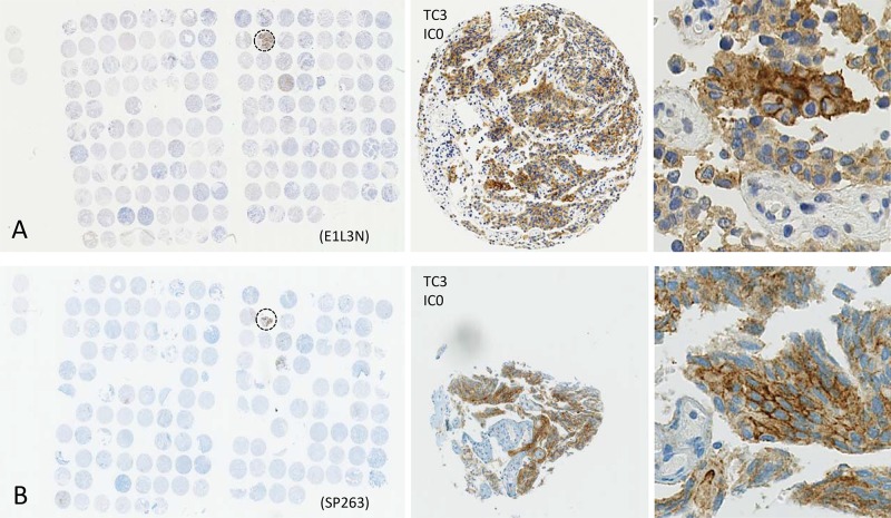
Figure 2.
Comparison of PD-L1 staining results for two different monoclonal anti-PD-L1 antibodies using tissue microarrays of CRPC specimens (panel (A) E1L3N, panel (B) SP263). Single tissue spots have a diameter of 0.6 mm. Zoomed versions, showing positive membranous PD-L1 immunoreactivity in a positive CRCP patient (dashed circle).

