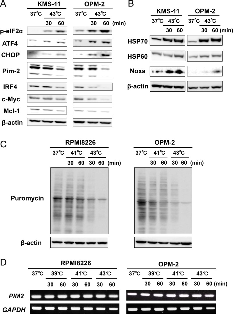
Figure 2. Hyperthermia induces ER stress along with the downregulation of IRF4, Pim-2, c-Myc and Mcl-1 in MM cells.
(A) KMS-11 and OPM-2 cells were cultured at 37 or 43°C for the indicated time periods. The cells were harvested, and the protein levels of phosphorylated eIF2α (p-eIF2α), ATF4, CHOP, IRF4, Pim-2, c-Myc and Mcl-1 were analyzed by Western blotting. β-actin was used as a protein loading control. (B) KMS-11 and OPM-2 cells were cultured at 37 or 43°C for the indicated time periods. The cells were harvested, and the protein levels of HSP70, HSP60 and Noxa were analyzed by Western blotting. β-actin was used as a protein loading control. (C) Puromycin incorporation. RPMI8226 and OPM-2 cells were treated with hyperthermia at 41 or 43°C for the indicated time periods. Puromycin was added at 1 μM for the last 15 minutes of the hyperthermia. The cells were harvested, and their puromycin incorporation was examined by Western blot analysis. β-actin was blotted as a loading control. (D) RPMI8226 and OPM-2 cells were treated with hyperthermia at 39 or 41 or 43°C for the indicated time periods. Then, the cells were incubated at 37°C for 6 hours. PIM2 mRNA expression was determined by RT-PCR. GAPDH mRNA was used as an internal control.

