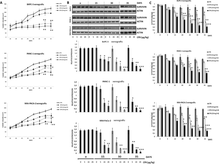
Figure 4. Pharmacodynamic analysis of CPX showed suppression of survivin levels in tumor of human pancreatic xenograft mice as well as inhibition of tumor growth, in a concetration- and time-dependent manner.
Female SCID mice were inoculated subcutaneously with BxPC-3 (3 × 10⁶ cells/mouse) or PANC- 1 (3 × 10⁶ cells/mouse) or MIA PaCa-2 (3 × 10⁶ cells/mouse) tumor cells. When local tumors were established, mice were randomly subdivided into groups after daily single oral gavage of CPX at concentration 5 mg/kg, 15 mg/kg, and 25mg/kg. Tumor tissues were resected on 0, 5th, 15th, 30th day after the daily administration of CPX. Tumor tissues were also resected on day 35th day (5 days after stopping the administration of CPX). (A) The tumor volumes within each cell line group were calculated (thrice/per week) after oral gavage of CPX at 5, 15 and 25 mg/kg daily. The mean ± SD (n = 6) data on tumor volumes are presented in the indicated graph *Significant difference (P < 0.05), **Significant difference (P < 0.01) versus controls (untreated) per time point. (B) Western blot analysis was followed for detecting survivin protein levels in tumor tissues (upper panel). The densitometric quantification of survivin reflects average values ± SD (n = 3) of at least 3 independent experiments. *p < 0.05 **P < 0.01 ***P < 0.001 vs. Control group of the indicated time point (lower panel). (C) Survivin levels as measured by ELISA after increasing doses of CPX at 5, 15, 25 mg/kg (n = 3 per time point). Survivin reflects average values ± SEM of at least 3 independent experiments. *p < 0.05 **P < 0.01 ***P < 0.001 vs. Control group of the indicated time point.

