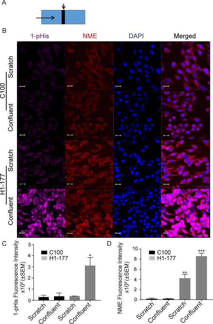
Figure 6. MDA-MB-435 migrating cells have a low 1-phosphohistidine level that does not correlate with total NME level.
A. Vector (C100) and NME1 (H1-177) overexpressing MDA-MB-435 cells were plated in chamber slides and treated as described in the legend for Figure 5A. B. After 24 hrs cells were fixed in PFA and immunofluorescence was performed for 1-pHis and NME1/2. Immunofluorescence staining was quantitated as total fluorescence intensity of 1-phosphohistidine (1-pHis) C. and total NME D. Both vector (C100) and NME overexpressing cells (H1-177) in the scratch area did not exhibit strong 1-pHis positivity. Nuclei were visualized using DAPI (Blue). Images were captured at 65x magnification in the scratch and confluent areas. Student's t-test was performed between Vector-Scratch vs NME-Scratch and Vector-Confluent vs NME-Confluent.

