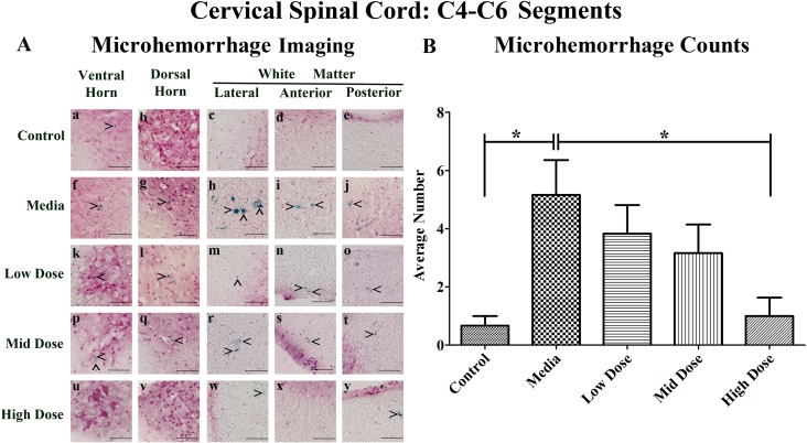
Figure 1. Distribution of microhemorrhages within the cervical (C4-C6) spinal cord of G93A mice after cell transplantation.
Tissue sections from the C4-C6 enlargement segment of the cervical spinal cord of 17 week old G93A mice were stained with Perl's Prussian blue to reveal ferric iron deposits as markers of microhemorrhages (blue). The tissue was counterstained with nuclear fast red (nuclei - red, cytoplasm - pink). (A) Numerous microhemorrhages (^) were detected within the parenchyma of the ventral and dorsal horns, lateral, anterior, and posterior white matter of the cervical spinal cord of ALS mice receiving media (f-j), low (k-o), or mid (p-t) cell dose. No microhemorrhages were seen in ventral or dorsal horns (u, v) and anterior white matter (x) of the high cell-dose mice. However, some small microhemorrhages were observed in the lateral white matter (w) and the posterior white matter (y) in these mice. A small iron deposit was detected in the ventral horn (a) of control mice. Yet, microhemorrhages were not detected in dorsal horn (b), or white matter (c-e) in these animals. In 50% of the high cell-dose mice, no microhemorrhages were observed. Scale bar is 50 μm. (B) Quantitative analysis of microhemorrhage distribution in the cervical spinal cord of the control and G93A mice. Media-treated mice showed a significantly (*p<0.05) higher number of microhemorrhages vs. controls. Microhemorrhages decreased inversely with cell dose, reaching significance with high cell-dose vs. media-treated mice (*p<0.05).

