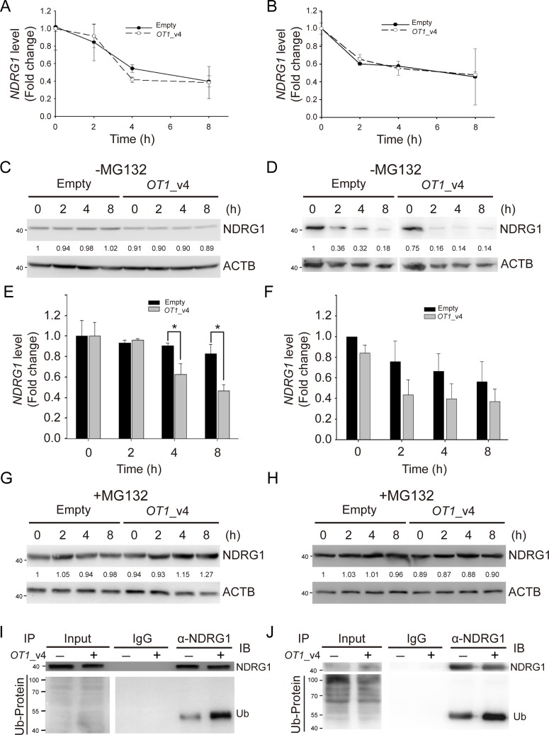
Figure 5. NDRG1-OT1_v4 promotes NDRG1 degradation via ubiquitination.
(A and B) Temporal profile of NDRG1 in NDRG1-OT1_v4-overexpressed MCF-7 (A) and SKBR3 (B) cells treated with actinomycin D (10 μg/mL). NDRG1 mRNA was measured by quantitative RT-PCR. Internal control: 18s. (C and D) Western blot of NDRG1 in NDRG1-OT1_v4-overexpressed MCF-7 (C) and SKBR3 (D) cells treated with cycloheximide (10 μg/mL). (E and F) Quantification of NDRG1 in (C&D). The results are the means ± SDs. *, P <0.05. (G and H) Western blot of NDRG1 in NDRG1-OT1_v4-overexpressed MCF-7 (G) and SKBR3 (H) cells treated with cycloheximide (10 μg/mL) and MG132 (20 μg/mL). (I and J) Co-immunoprecipitation of NDRG1 and ubiquitin in MCF-7 (I) and SKBR3 (J) cells overexpressing NDRG1-OT1_v4 in the presence of MG132 (20 μg/mL). Cells were immunoprecipitated with NDRG1 antibody, followed by western blotting. Data were repeated at least 3 times.

