
Figure 1. A higher proportion of immature vasculature correlates with enhanced survival rates of mCRC patients following bevacizumab treatment.
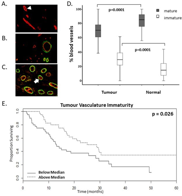
(A-C). Representative images of dual immunofluorescent staining for FVIII (red) and α-smooth muscle actin (αSMA) (green) indicative of pericyte recruitment to mature vessels. Varying levels of vasculature maturity was observed from (A.) low, (B.) moderate and (C.) high levels of vasculature maturity in the resected colorectal cancer tumour microenvironment. Arrowhead highlights an immature vessel; arrow highlights a mature vessel which has recruited pericytes. D. Graph shows the proportion of mature and immature vasculature in tumour tissue compared to matched normal tissue. There is a lower percentage of mature vasculature and a higher percentage of immature vasculature in tumour tissue compared to normal tissue (p values=0.0001, n=80). E. Kaplan-Meier survival curves of above (dotted line) and below (continuous line) median levels of percentage immature vasculature (% IMM) prior to commencing bevacizumab treatment versus overall survival in months (p value=0.026, n=80).