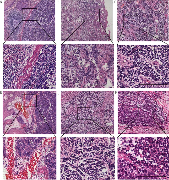
Figure 2.
(A) The boundary between NEC and squamous cell carcinoma is clear and lymphocytes infiltrating into lymphoid follicles. (B) NEC cells were mixed with esophageal squamous carcinoma cells without a border. Tumor stroma was infused with blood vessels in most cases with capillaries (C) and venules (D). Lymph node metastasis could be found in 24 cases (E, F). Hematoxylin-eosin stain in A-D.(Scale bar: 100 μm)

