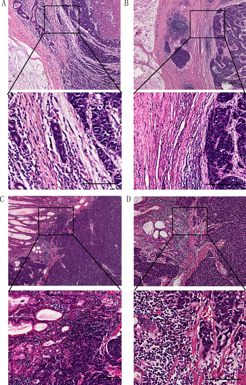
Figure 3. In gastric cardia NECs, some cases were mixed with mucinous adenocarcinoma, and with a clear border (A, B).

Some were mixed with adenocarcinoma, and with a broken boundary (C, D).(Scale bar: 100 μm)

Some were mixed with adenocarcinoma, and with a broken boundary (C, D).(Scale bar: 100 μm)