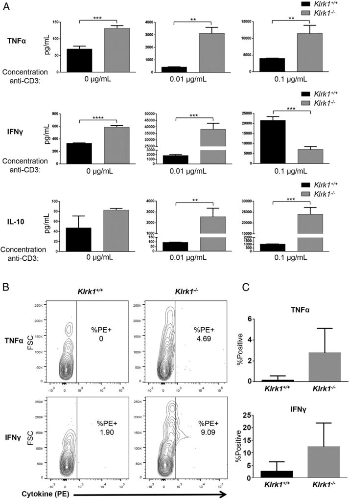
FIGURE 4. Altered cytokine production by Klrk1−/− NOD CTL.
(A) Secretion of TNF-α, IFN-γ, and IL-10 (mean ± STD) by in vitro–generated Klrk1−/− and wild-type CTL stimulated with the indicated concentrations of anti-CD3ε Ab. Data are representative of at least six independent experiments. (B) Representative flow cytometry plots showing intracellular staining of TNF-α and IFN-γ in Klrk1−/− and wild-type NOD CD8+ T cells 1 wk after cotransfer into a wild-type NOD adoptive transfer recipient mouse. (C) Combined results (mean ± SEM) from transfers into eight mice from three independent experiments showing the percent TNF-α or IFN-γ positive Klrk1−/− and wild-type NOD CD8+ T cells 1 wk after cotransfer into wild-type NOD adoptive transfer recipient mice. **p ≤ 0.01, ***p ≤ 0.001, ****p ≤ 0.0001 in two-tailed unpaired Mann–Whitney U test.

