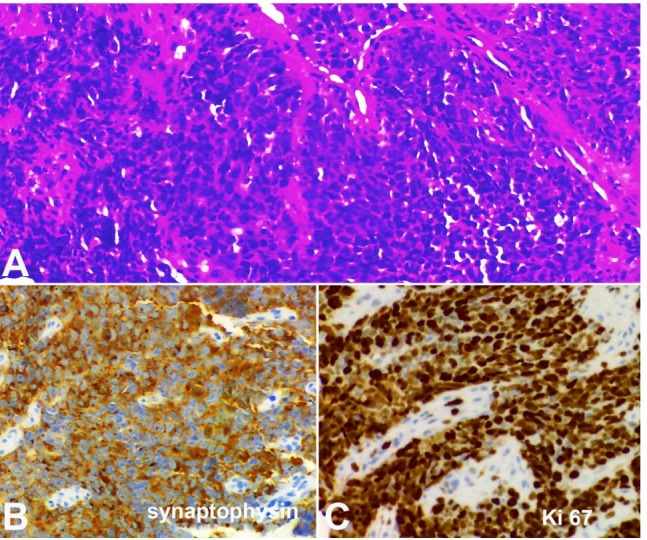
Figure 2. Photomicrography of the gastric biopsy. A – Nests of a poorly differentiated carcinoma stained with hematoxylin-eosin; B – Diffuse cytoplasmic synaptophysin immunostaining; C – High cell proliferation index, showing 90% of Ki-67 positive cells.

